Figure 1.
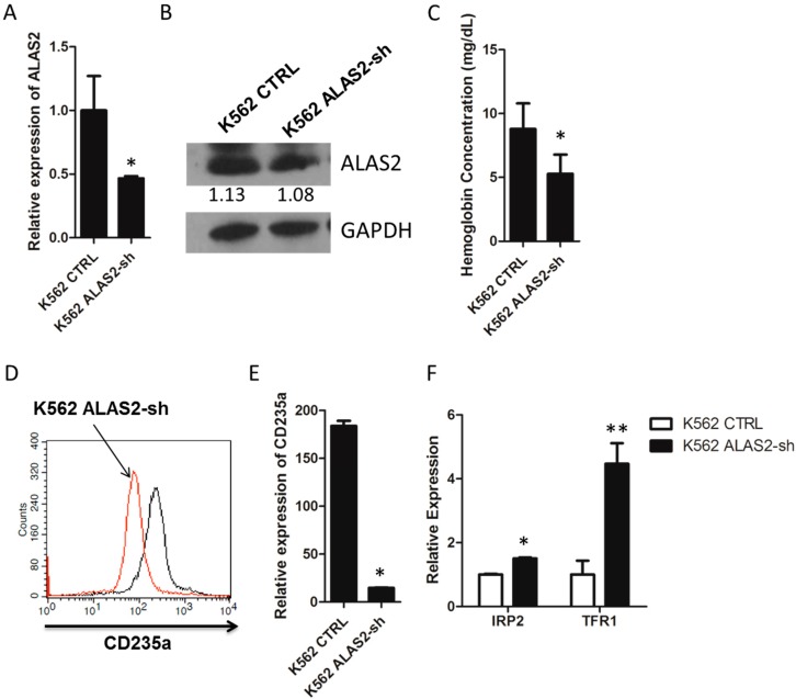
(A) Relative mRNA expression of ALAS2 in K562 control and K562 ALAS2-sh cells using quantitative real-time PCR; (B) Representative Western blots showing ALAS2 expression in K562 control and K562 ALAS2-sh cells. Quantification normalized to GAPDH by densitometry using ImageJ was provided; (C) Hemoglobin concentration of K562 control and K562 ALAS2-sh cells. Cell surface expression of CD235a (D) and mRNA levels (E) of K562 control (black line in D) and K562 ALAS2-sh using FACS and quantitative real-time PCR, respectively; (F) Relative mRNA expression of IRP2 and TFR1 in K562 control and K562 ALAS2-sh cells using quantitative real-time PCR. All relative mRNA expression using quantitative real-time PCR was normalized to glyceraldehyde-3-phosphate dehydrogenase (GAPDH) expression. Asterisks indicate that differences between samples were statistically significant according to an independent-sample t-test, * p < 0.05, ** p < 0.01.

