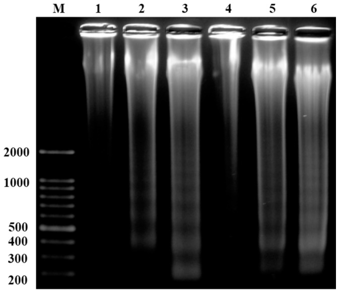
Figure 4.
Linalool administered to HeLa cells at concentrations of 6.48 μM (2) and 12.96 μM (3) and activated for 6 h. Linalool administered to U937 cells at concentrations of 1.94 μM (5) and 3.24 μM (6) and activated for 6 h. The DNA damage following cell apoptosis was determined via agarose gel electrophoresis. Columns 1 and 4 show the control group, and M shows the DNA marker group.

