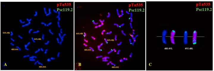
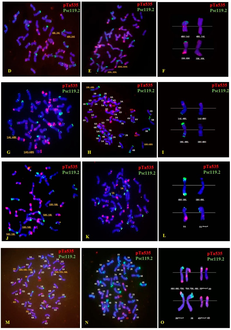
Figure 3.
Fluorescence in situ hybridization (FISH) performed usingpTa535 (red) and pSc119.2 (green) as probes for chromosomes with structural changes and mutants in the M1 generation. (A,B) 4B.6V translocation; (D) 2A.6B translocation; (E) 2D.3D translocation; (G) 1A.6B translocation; (H) 1B.6B translocation, and 6D trisome; (J) 5B.1B translocation; (K) 5A aberrations; (M) 5B.7D translocation, 3D and 6D aberrations, and 1D monosome; (N) 6D trisome. Images (C,F,I,L,O) show enlargements of the FISH pattern of chromosomes involved in the structural changes. Chromosomes were counterstained with 4′-6-diamidino-2-phenylindole (blue).


