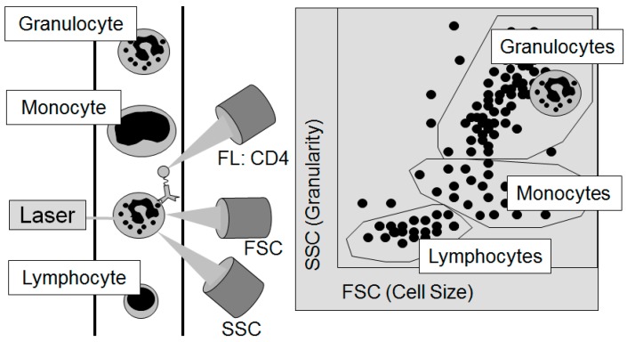
Figure 3.
The principle of flow cytometry to differentiate the individual cellular components of the blood: The Forward Scatter (FSC) measures the acute-angular diffraction of the light and depends on the cell’s volume. The Sideward Scatter (SSC) measures the rectangular diffraction of the light and depends on the granularity of the cell, the size and structure of its core, and the amount of vesicles contained in it. With the help of FSC and SSC, the individual cells contained in the blood can be differentiated. For further sub-differentiation, e.g., by the sub-type of cell or its functional status, marker molecules on the surface of the cells, the so-called cluster of differentiation (CD), are used. In this figure, a granulocyte passes the laser light (left part of the figure). After sorting by cell size and granularity according to the light detected by the FSC and the SSC as displayed in the right part of the figure, the granulocyte could then be further analyzed for the frequency of CD4 on its surface by the signal of fluorescent (FL) antibodies against CD4.

