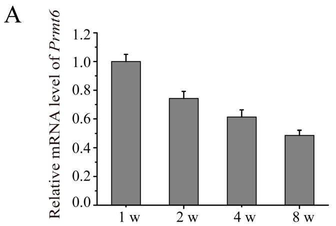
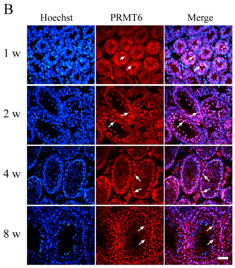
Figure 2.
The expression of Prmt6 mRNA and PRMT6 protein during mouse testes development. (A) RT-qPCR analysis was used to detect Prmt6 mRNA in developing postnatal mouse testes. The relative expression of Prmt6 at different stages of testes development was compared to one week (w). Gapdh was used as an internal control. Data are expressed as the mean ± SD (n = 5); (B) Immunofluorescence analysis was used to determine the localization of PRMT6 in mouse testes using a PRMT6 antibody (red). The nuclei of cells were labeled with Hoechst 33342 (blue). Purple represented the merging color of red and blue. Arrow: spermatogonia or spermatocytes. Bar = 50 μm.


