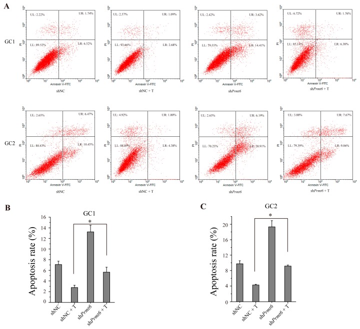
Figure 7.
Germ cell apoptosis was promoted by transfecting GC-1 and GC-2 cells with shPrmt6 and treated with testosterone (T). (A) Representative graphs of GC-1 and GC-2 cell apoptosis as analyzed by flow cytometry. LR (lower right section of the graphs) which was Annexin V-FITC positive and PI (propidium iodide) negative represented the percentage of apoptosis cells; (B,C) Statistical analysis of the cell apoptosis rate of GC-1 and GC-2 cells. All of the experiments were repeated at least three times. Data are expressed as the mean ± SD. * p < 0.05.

