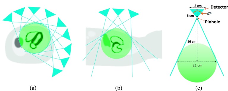
FIG. 1.
Design of the dedicated cardiac multiple pinhole SPECT scanner. (a) This diagram illustrates how nine pinholes surrounding a NCAT phantom are all simultaneously imaging the heart in transaxial view. The green sphere presents the FOV of the scanner. The pinhole apertures are distributed on a cylindrical surface with a radius of 16 cm. (b) The pinhole collimators shown in longitudinal axis. (c) Profile of a single detector on a pinhole.

