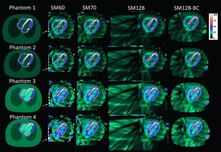
FIG. 4.
The central transaxial slices of the reconstructed images using different SM of the noiseless NCAT phantom. All the images were reconstructed with 100 iterations. The yellow dashed arrows denote the artifacts on the edge of the FOV. The white solid arrows denote the artifacts in the blood pool.

