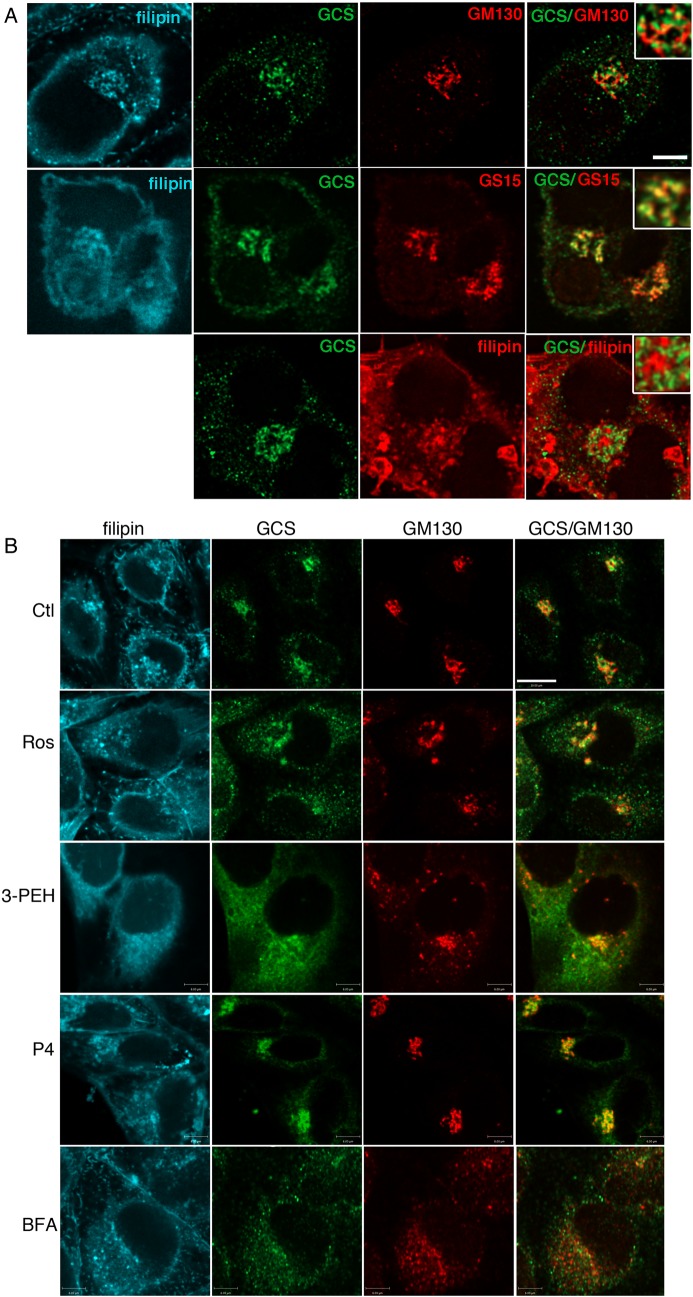
Fig. 6.
Golgi GCS is relocalized after statin or 3-PEHPC treatment. (A) After permeablization with 50 µg/mL filipin cells were double-labeled with rabbit anti-GCS 1.2 antiserum and mAb GM130 (cis-Golgi) or mAb GS15 (medial Golgi). GCS was localized in punctate Golgi structures partially overlapping with GS15, and proximal but largely distinct from GM130 and filipin-labeled TGN (pseudo-colored red in the bottom panel for clarity). Scale bar = 6 µm. (B) ACHN cells were treated 48 h with vehicle, 10 µM rosuvastatin, 1 mM 3-PEHPC, 2 µM P4 or 30 min with 10 µM BFA, prior to fixation and labeling. After statin or 3-PEHPC treatment, GCS became more widely distributed, the TGN cholesterol pool (but not PM cholesterol) was absent, anti-GM130-labeled dispersed Golgi “mini-stacks”. Scale bar = 10 µm.

