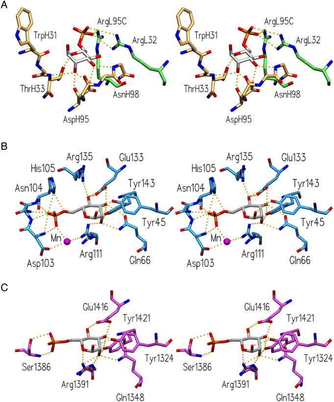
Fig. 5.
Comparison of Man6P-binding sites of Fv M6P-1 and bovine MPR. Stereoviews of the binding sites of (A) Fv M6P-1 (light chain green, heavy chain tan), (B) the CRD of MPR46 (pdb code 1M6P; Roberts et al. 1998) and (C) the CRD formed by the N-terminal domains 1–3 of MPR300 (pdb code 1SYO; Olson et al. 2004), showing hydrogen bonds and salt bridges as dashed yellow spheres.

