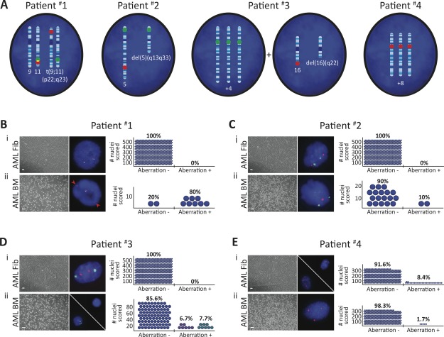
Figure 1.
The majority of acute myeloid leukemia (AML) Fibs are devoid of leukemia-associated aberration. (A): Schematics illustrating patient-specific leukemic aberration(s) identified in AML blast nuclei. Fluorescence in situ hybridization (FISH) probe hybridization regions are indicated (green/red) on affected chromosomes. (B–E): FISH performed in AML patient-derived (i) Fibs and (ii) bone marrow (BM) mononuclear cells (scale bars represent 100 µm); n = 1 per AML patient. Aberrations were detected in each patient AML BM, and a population of patient #4 AML Fibs. Red arrows denote probe separation associated with translocation in patient #1 AML BM. Adjacent plots depict the frequency of detection of patient-specific, leukemia-associated aberration; blue circles represent number of nuclei analyzed. Blue circles with either one red dot or three green dots represent del(16)(q22) and +4 events in patient #3 AML BM, respectively; aberrations were never detected in the same nuclei. 500 nuclei were analyzed to exclude 1% genetic mosaicism in AML patient #1–3 Fibs with 99% confidence 41.

