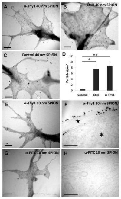
Figure 2. Functionalized SPIONs attach to RGC membranes.
Transmitted electron microscope (TEM) images of RGC growth cones treated with (a) 40 nm anti-Thy1 SPIONs and (b) 40 nm anti-CtxB SPIONs for 30 minutes show abundant attachment of functionalized SPIONs to filopodia and other parts of the growth cone. (c) In contrast, very few non-functionalized 40 nm SPIONs are found at growth cone structures. (d) Quantification of number per surface unit of 40 nm iron core SPIONs functionalized with different moieties. Results are expressed as mean ± SEM of at least 4 growth cones in two independent experiments. Significance differences between functionalized and control non-functionalized SPIONs were tested using an unpaired Student’s t-test. *p<0.05, **p<0.01. (e) TEM images of RGC growth cones incubated with 10 nm iron core anti-Thy1 SPIONs for 30 minutes show abundant SPION attachment. (f) TEM images of RGC microtome sections reveal that 10 nm iron core anti-Thy1 SPIONs remain on the outside membrane of the cell body (*) and neurite (star). (g) In contrast, 10 nm iron core anti-FITC SPIONs were not found in RGC growth cones nor in (h) RGC microtome sections. Scale bar 1 μm in a, b, c, e and g and 200 nm in f and h.

