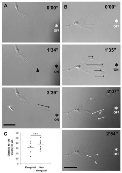
Figure 3. Filopodia elongation and retraction in response to On/Off magnetic field.
(a) Filopodium from a cultured RGC treated with anti-thy1 SPION elongated (black arrow) towards the tip of the applied electromagnet (asterisk) during the first several minutes. An accumulation of membrane material in response to the magnetic field was observed at the tip of the filopodium (arrowhead) preceding elongation. (b) Similarly, in RGCs treated with CtxB SPIONs, several filopodia elongated (black arrows) in response to an applied magnetic field (ON). These filopodia rapidly retracted (white arrows) when the magnet was turned off (OFF). Scale bar 10 μm in a and b. (c) Distances from the end of each filopodium to the tip of the electromagnet were measured before elongation. From the total filopodia of a growth cone, the closest ones to the electromagnet tip were more likely to elongate towards the magnet (***p<0.001 by unpaired Student’s t-test; n=6, 13; distances from tip of each filopodium to the magnet were normalized to the average distance of filopodia that elongated in that growth cone).

