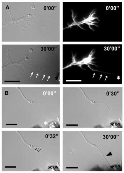
Figure 4. Filopodial actin and growth cone responses to elongation.
(a) Thirty minutes after elongating a Thy1-SPION-treated filopodium to a PDL- and laminin-coated electromagnet tip (asterisk), the RGC growth cone did not change its position. Expression of RFP-LifeAct peptide in RGCs allowed visualization of F-actin filaments inside the elongated filopodium (white arrows). (b) A filopodium was elongated using CtxB-SPION, and anchored to the PDL-/laminin-coated electromagnet tip (asterisk). Engorgement of the filopodium after 30 minutes was observed (black arrowhead). The originating segment of the filopodium relocated to a new position (black arrows) in response to mechanical tension. Scale bar 10 μm in a and b.

