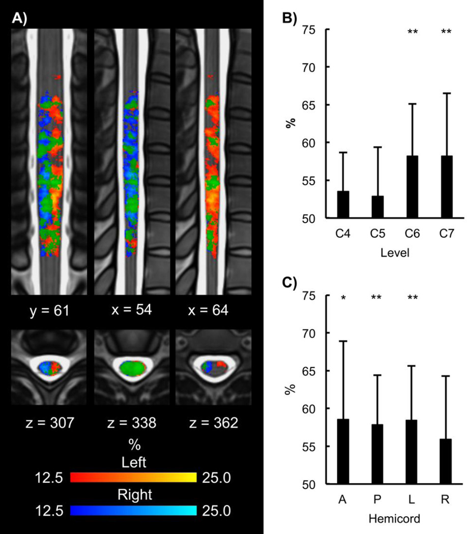
Figure 6.
A) Across subject trialwise consistency maps (1 coronal, 2 sagittal, and 3 axial slices) for the left (yellow-red) and right (blue-light blue) contrasts (overlap in green). The consistency maps show the percentage across all trials and all subjects that a voxel was active (Zscore > 1.65). Overall, the consistency maps demonstrate lateralization of the activity to the hemicord ipsilateral to the task at the trial level. B) Percent accuracy of the trialwise multi-voxel pattern analysis (MVPA) at each vertebral level is shown. MVPA was able to decode the left and right contrasts at the C6 and C7 vertebral levels better than chance. C) Trialwise MVPA was then performed across the C6 to C7 vertebral levels in the anterior, posterior, left, and right hemicords. At C6 to C7, MVPA was able to decode the left and right contrasts in the anterior, posterior, and left hemicords. *p < 0.05 and **p < 0.01. A = anterior, P = posterior, L = left, and R = right.

