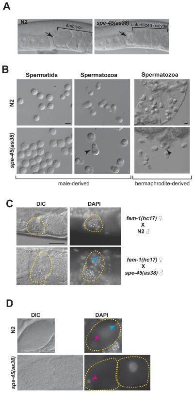
Figure 2. Despite Normal Sperm Morphology and Migratory Behavior, spe-45 Mutant Sperm Cannot Fertilize Oocytes.
(A) DIC images of wild-type and spe-45(as38) reproductive tract. Black arrow indicates spermatheca containing sperm. Uterus is located at the right side of spermatheca; ovary is located at the left side of spermatheca. Scale bar: 20μm.
(B) Spermatids and pronase-activated spermatozoa of N2 and spe-45(as38) males (Left panels). Spermatozoa from N2 and spe-45(as38) hermaphrodites (Right panels). Arrow head indicates pseudopod of sperm. Scale bar: 5μm.
(C) DAPI staining of the fem-1(hc17) females mated with either N2 males or spe-45(as38) males. Yellow, dotted region shows spermatheca regions are indicated by. Blue arrow head indicates an example of sperm DNA. Scale bar: 20μm.
(D) DAPI staining of N2 and spe-45(as38). Blue arrowhead indicates the compacted sperm chromatin mass in a newly fertilized oocyte; the pink arrowheads indicate the meiotic oocyte chromosomes in a newly fertilized oocyte. The white arrowhead indicates the chromosomes of an unfertilized oocyte that have undergone endomitotic replication. See related Figure S2. Scale bar: 10μm

