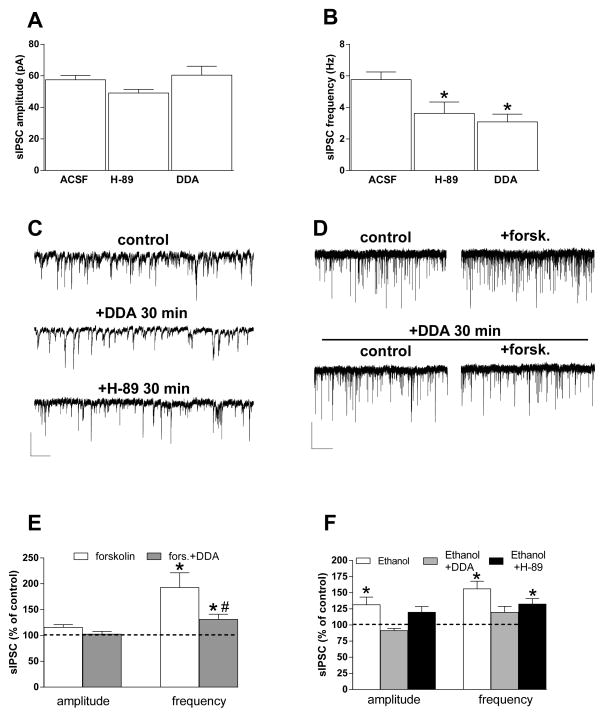
Fig. 2. Roles of adenylyl cyclase and protein kinase A in ethanol potentiation of sIPSCs.
A, B) Bar graphs showing the effect of 30-min incubation of slices with the PKA inhibitor H-89 (10 μM) or the AC inhibitor DDA (10 μM). Neither compound altered sIPSC amplitude (A), but both produced a decrease in sIPSC frequency (B) (n = 54, 11, 21) (*p < 0.05 vs. aCSF unpaired t test. C) Representative traces obtained from different neurons incubated in normal aCSF, or after 30 min of H-89 or DDA incubation. Scale bar = 50 pA, 10 sec. D, E) Forskolin (20 μM) increased sIPSC frequency, and this effect was dramatically decreased in slices incubated for 30 min with DDA. Representative current traces in D show forskolin effects on sIPSCs in the absence (top) and presence (bottom) of DDA. Scale bar = 50 pA, 10 sec. Bar graphs in E show the forskolin and forskolin + DDA effects on sIPSC amplitude and frequency (n = 5, 3) (*p < 0.05 vs. baseline paired t test; #p < 0.05 vs. forskolin, unpaired t test). F) The AC inhibitor DDA decreases ethanol potentiation of the amplitude and frequency of sIPSCs recorded from BLA-projecting neurons, while the PKA inhibitor H-89 did not significantly alter ethanol actions (n = 27, 7, 5) (*p < 0.05 vs. baseline, paired t test).

