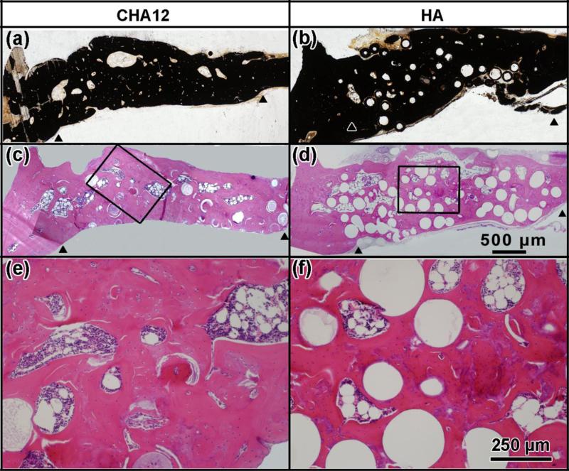
Fig. 6.
Transmitted light images of (a, b) von Kossa and (c, d) H&E stained sections of rat calvarial defects implanted with hollow CHA12 and HA microspheres loaded with BMP2 at 12 weeks postimplantation; (e, f) higher magnification images of the boxed areas in c, d. (Arrowheads: edges of host bone)

