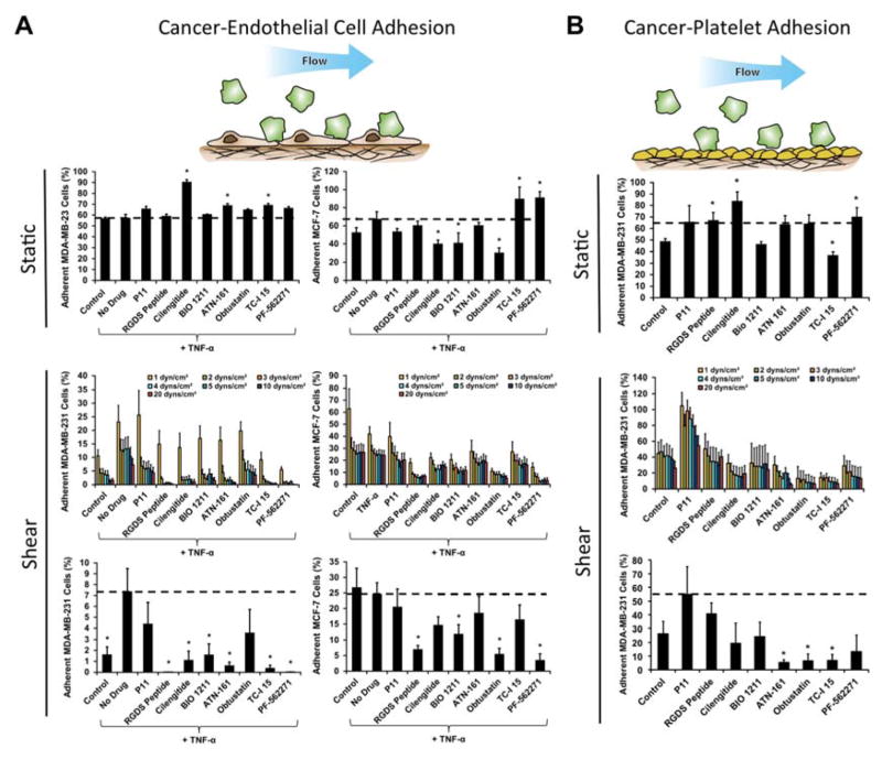
Fig 4.
Screening of integrin inhibitors under static and flow conditions using a cancer cell de-adhesion assay. (A) Plots of relative adhesion of MDA-MB-231 or MCF-7 breast cancer cell lines under static conditions, the adhesion/detachment assay under 0.5 dynes/cm2 shear stress in the presence of integrin inhibitors, and the remaining adherent cells after 20 dynes/cm2 shear stress had been applied. (B) Plots of relative adhesion of MDA-MB-231 cells to immobilized platelets under static and flow. *p < 0.05 versus TNF-α treated cells.

