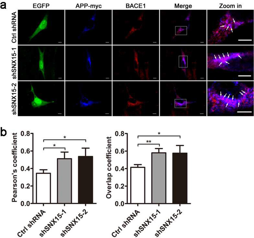
Fig. 5.
Downregulation of SNX15 increases the colocalization of APP and BACE1. a HT22 cells co-transfected with APP-myc and Ctrl shRNA, shSNX15-1 or shSNX15-2 (EGFP, shown in green), were immunostained with anti-myc primary antibody followed by fluorescence-conjugated secondary antibody (in blue) and with anti-BACE1 primary antibody followed by fluorescence-conjugated secondary antibody (in red), and then observed under the Olympus FV1000 confocal microscope. Enlarged areas in the merged images on the right show colocalization of APP and BACE1 in purple (white arrows). Scale bars: 5 µm. b Both Pearson's coefficient and overlap coefficient were measured by the Olympus imaging analysis software to indicate colocalization of APP and BACE1. At least ten randomly selected neurons from each treatment were measured for comparison. *p<0.05, **p<0.01.

