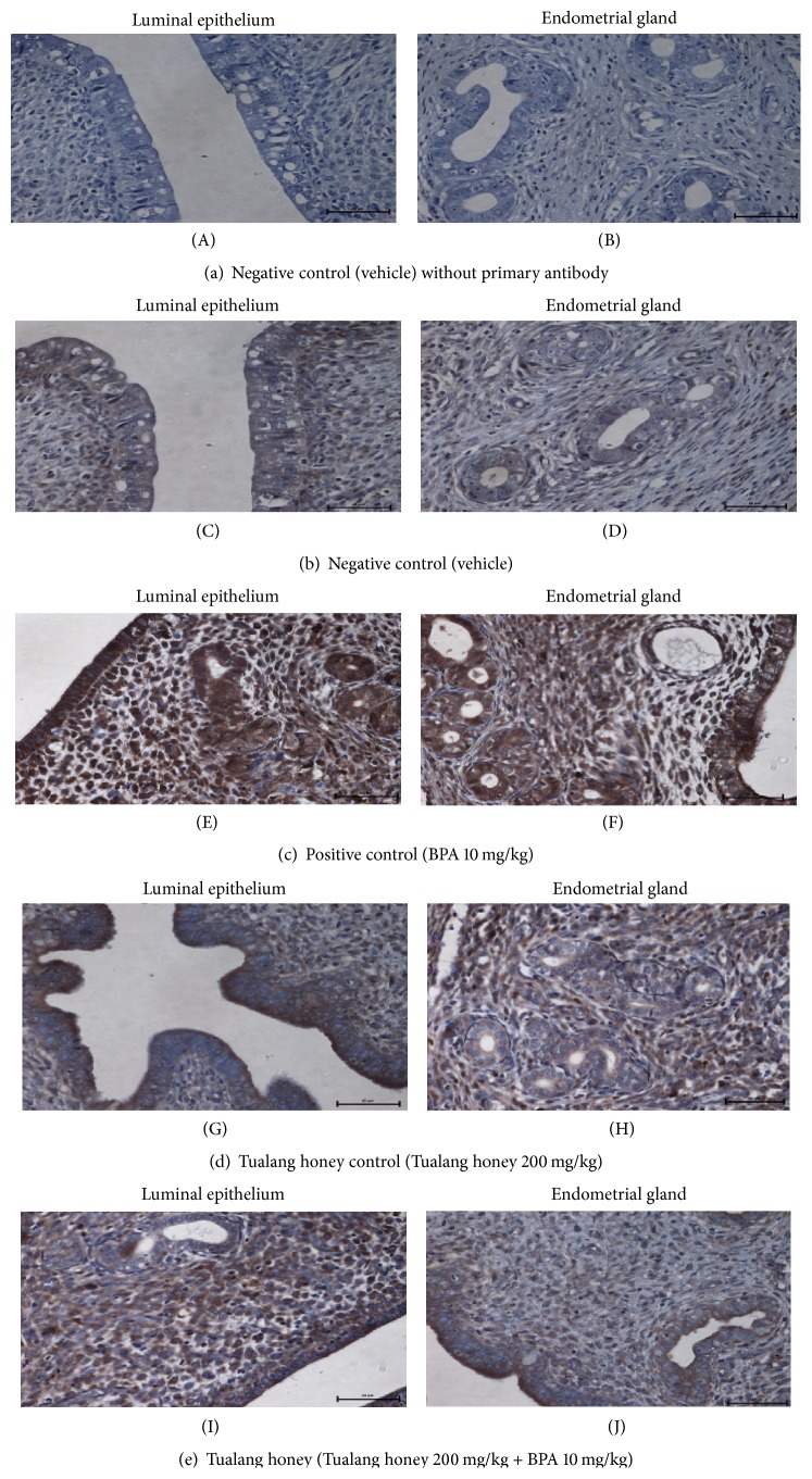
Figure 6.
Representative sections of uteri showing the immunohistological localization of ERβ in all experimental groups (×40). The most pronounced immunostaining intensity was observed in BPA-exposed rats ((E), (F)) (PC group). Comparable immunostaining intensities were observed in NC ((C), (D)), THC ((G), (H)), and TH ((I), (J)) groups.

