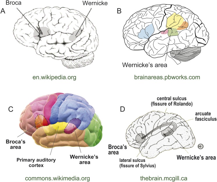
Figure 1. Current depictions of the Wernicke area.
A representative sample of Internet images depict the Wernicke area, found using a Google search for “Wernicke's area images.” All highlight the posterior superior temporal gyrus, with variable extension into the posterior supramarginal gyrus and occasionally the angular gyrus. Web locations are shown in green below each image. A–C are in the public domain; D is reproduced with permission from the Web page manager.

