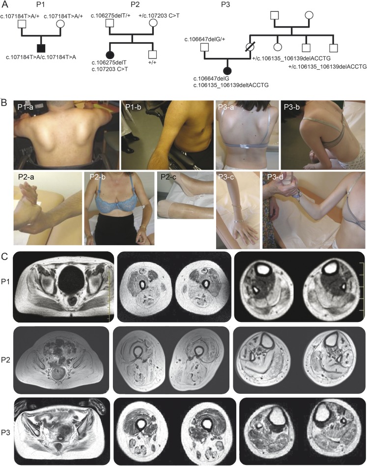
Figure 1. Pedigree of the families and clinical presentation and imaging of the patients.
(A) Pedigree of the families and clinical presentation and imaging of the patients. Filled-in symbols indicate the affected individual. Empty symbols indicate unaffected individuals. Numbers indicate individuals whose DNA was used for this study. The mutations identified are indicated for each tested individual. (B) Photographs of patients showing prominent contractures. Patient 1 (P1): P1-a: severe upper girdle involvement with scapular winging; P1-b: arm atrophy and elbow contractures. Patient 2 (P2): P2-a: finger flexor contractures; P2-b: severe proximal involvement and elbow contractures; P2-c: severe ankle contracture. Patient 3 (P3): P3-a: severe upper involvement with scapular winging; P3-b: rigid spine; P3-c, P3-d: finger and elbow contractures. (C) Muscle MRI of the patients at 3 different levels (axial T2 images through pelvis, thigh, and calf). P1 at 23 years: diffuse fatty degeneration in gluteal muscles with partial sparing of the sartorius and severe involvement of other femoral muscles and of the posterior and lateral compartments as well as tibialis anterior of the lower legs. P2 at 52 years: severe and diffuse involvement with fatty replacement of all muscles. P3 at 20 years: diffuse moderate fatty degenerative changes in all muscle groups with atrophy more marked in the hamstrings.

