Figure 1.

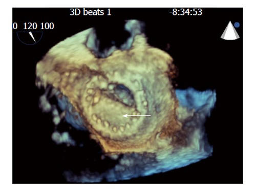
Still frames of 3-dimensional transesophogeal echocardiographic rendering of the mechanical bi-leaflet mitral valve as visualized from the left atrial perspective during diastole showing fixed mitral leaflet (arrow).

Still frames of 3-dimensional transesophogeal echocardiographic rendering of the mechanical bi-leaflet mitral valve as visualized from the left atrial perspective during diastole showing fixed mitral leaflet (arrow).