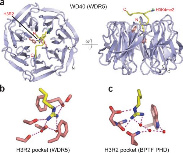
Figure 5.
Readout of an unmodified arginine by the WD40 repeat of WDR5. (a) Top view (left) and side view (right) of WDR5 in complex with H3K4me2 peptide (PDB 2H6N). H3R2 and H3K4me2 are in stick representation. H3R2 is deeply buried in the central cavity of WDR5, whereas H3K4me2 is presented by WDR5 on the protein surface for further methylation. (b,c) Details of H3R2 readout by WDR5 in a cavity-insertion recognition mode (b) and by BPTF PHD finger in a surface-groove recognition mode (c).

