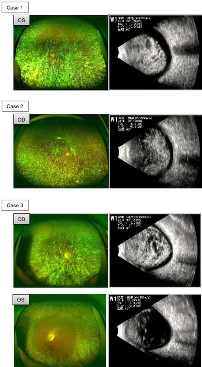
Figure 1.
Preoperative fundus photographs obtained by Optos200 Tx (Optos left images) and ultrasound B scan images (right images) in three cases. Owing to the dense opacity of the vitreous associated with the asteroid hyalosis, details of the fundus were unclear in the left eye in case 1, the right eye in case 2 and the right eye in case 3. Ultrasound B scan showed high acoustic echo in almost the entire vitreous cavity in these three eyes. In the left eye of case 3, which had mild opacity of the vitreous due to asteroid hyalosis, the ultrasound B scan showed a mild acoustic echo in the vitreous.

