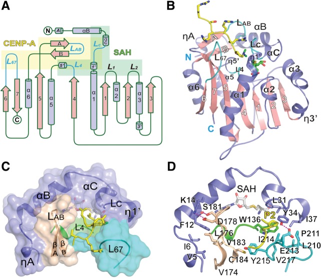
Figure 1.

Overall structure of the NRMT1–SAH–CENP-A ternary complex. (A) Topology of NRMT1. (Slate blue) Helices; (pink) β strands. CENP-A- and SAH-binding motifs are shaded yellow and green, respectively. CENP-A-interacting loops (LC, L4, LAB, and L67) are highlighted in cyan. (B) NRMT1 in complex with SAH and dimethylated CENP-A peptide. The CENP-A peptide and SAH are depicted as sticks. Color coding is the same as in A. (C) Composition of the CENP-A platform. Several segments of NRMT1 constitute the CENP-A peptide-binding pocket, including N-terminal helices (slate blue), loop L4 (green), βAB hairpin (wheat), and loop L67 (cyan). (D) Interaction details that enable an intimate integration of the CENP-A platform. Key residues are shown as sticks and are color-coded in the background of cartoon.
