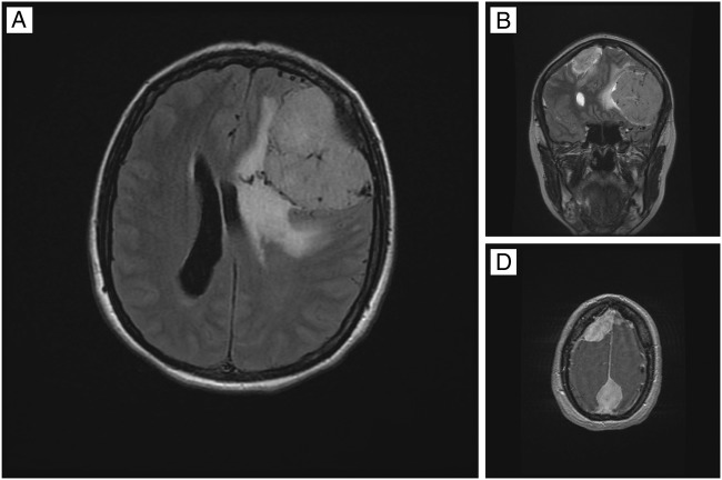
Figure 1.
MRIs indicating multiple brain meningiomas. (A) Brain MRI, axial plane, T2 fluid-attenuated inversion recovery (FLAIR) sequence: giant meningioma on the left frontal lobe with cerebral oedema and mass effect. (B) Brain MRI, coronal plane, T2 FLAIR sequence: meningioma on the left frontal lobe and a smaller meningioma on the right frontal lobe. (C) Brain MRI, axial plane, T2 FLAIR sequence: meningioma on the right frontal lobe and another meningioma on the parasagittal parietal lobe.

