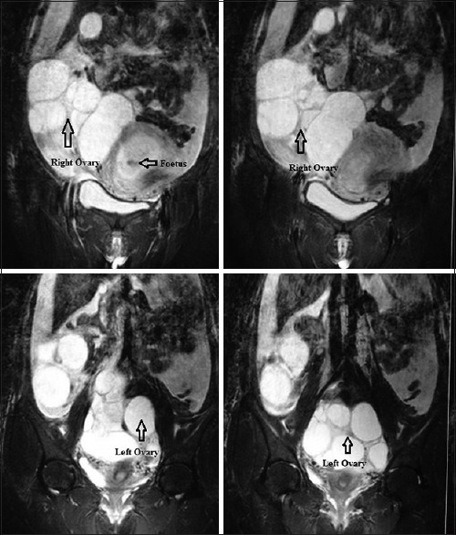
Figure 3.

T2-weighted magnetic resonance imaging pelvic coronal images show T2-hypo intense lesion in the intrauterine cavity suggestive of fetus. There are hyper intense multiple cysts seen without any mural nodule arising from both right (in right iliac fossa) and left ovary (in pouch of Douglas)
