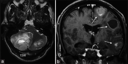
Figure 1.

Magnetic resonance imaging of the brain, (a) T2-weighted axial acquisition showing a large cerebellar tumor with edema and fourth ventricle compression. (b) T1-weighted following contrast administration is showing multiple metastatic lesions in the left cerebral hemisphere, solid, and cystic
