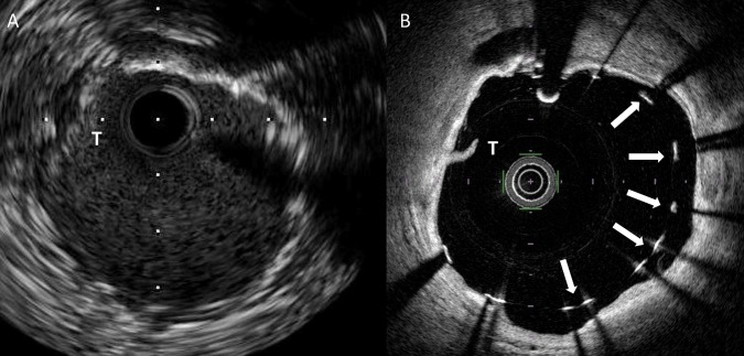
Figure 2.
(A) Matched IVUS and (B) FD-OCT cross-sectional images following coronary stent implantation in a patient treated with rotational atherectomy. An arc of calcification is visible in the vessel wall from the 12 o'clock position around to the 6 o'clock position. Malapposed stent struts are clearly visible in this area with FD-OCT (panel B, arrows) but the interface between stent strut and vessel wall is poorly delineated in the corresponding IVUS image (panel A). An area of thrombus adherent to the luminal surface of the stent is also visible (T). FD-OCT, frequency domain optical coherence tomography; IVUS, intravascular ultrasound.

