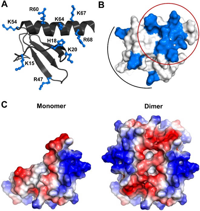
Figure 4. Distribution of basic residues that mediate heparin interactions.

(A) A schematic of CXCL8 monomer showing the distribution of basic residues arginine, lysine and histidine in ball and stick. (B) Surface plot of CXCL8 monomer with the GAG-binding residues shown in blue. The core residues that encompass the C-helix and N-loop are circled and the peripheral residues are highlighted by an arc. (C) Electrostatic surface plots of CXCL8 monomer and dimer.
