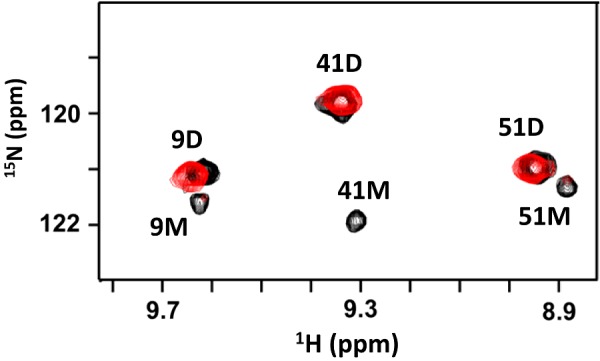
Figure 8. NMR HSQC spectra of CXCL8–dp26 complex.

A section of the 1H-15N HSQC spectra at pH 7.5 showing the overlay of WT CXCL8 (black) and WT CXCL8–dp26 complex (red). Dimer and monomer peaks are indicated by D and M respectively. The monomer peaks disappear on dp26 binding indicating tighter binding to the dimer. At pH 7.5 and 40 μM concentration, WT CXCL8 exists as both monomers (∼10%) and dimers. The final protein/dp26 molar ratio is 1:2.
