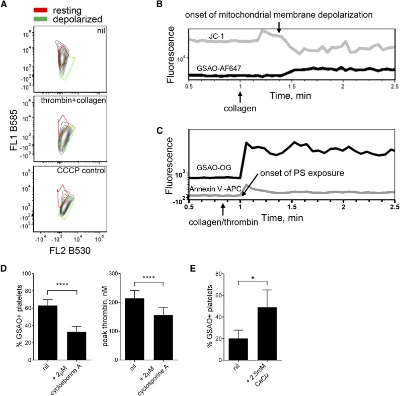
Figure 4.
GSAO marks platelets undergoing cyclophilin D-dependent necrosis. (A) Washed human platelets were untreated or stimulated with thrombin (0.1 U/mL) and collagen (5 µg/mL) or the mitochondrial membrane disrupter m-chlorophenylhydrazone (4 μM). Mitochondrial transmembrane potential was measured using the cationic dye, JC-1. The ratio of distribution of JC-1 between the mitochondria (red fluorescence) and cytosol (green fluorescence) reflects mitochondrial transmembrane potential. Results are representative of n ≥ 3 separate experiments. (B-C) The correlation between loss of platelet mitochondrial transmembrane potential (JC-1 loss of B585 fluorescence) or phosphatidylserine exposure (annexin V labeling) and labeling with GSAO-AF647 after collagen (5 µg/mL) or thrombin (0.1 U/mL) and collagen (5 µg/mL) stimulation was measured by time-lapse flow cytometry. (D) Washed human platelets were untreated or preincubated with the cyclophilin D inhibitor, cyclosporine A (CysA, 2 μM), for 15 minutes before stimulation with thrombin (0.1 U/mL) and collagen (5 µg/mL). Labeling with GSAO-AF647 was measured by flow cytometry and procoagulant potential assessed using the Calibrated Automated Thrombogram (n = 3-6; ****P < .0001). (E) Washed human platelets were preincubated with CaCl2 for 15 minutes and then untreated or stimulated with thrombin (0.1 U/mL) and collagen (5 µg/mL). Labeling with GSAO-AF647 was measured by flow cytometry (n = 8; *P < .05).

