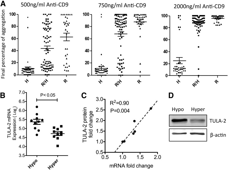
Figure 1.
Hyperresponders to FcγRIIA-mediated platelet activation have reduced TULA-2 expression compared with hyporesponders. (A) Platelets from 154 healthy human donors were activated by indicated dose of anti-human CD9. Final percentage of aggregation was used as the readout for reactivity. This population was further divided by genetic variation at the codon 131 of FCGR2A gene. Hyperresponders were defined as having >75% final aggregation at 750 ng/mL anti-CD9, whereas hyporesponders were defined as having <25% aggregation. (B) TULA-2 mRNA (UBASH3B) is differentially expressed between 10 top hyperresponders and 10 bottom hyporesponders. All donors were ranked based on the final percentage of aggregation (Student t test, P < .05). (C) TULA-2 protein levels were measured by western blot. The correlation between TULA-2 protein level and TULA-2 mRNA level was determined by Pearson correlation (R2 = 0.90). (D) Representative western blot of platelet TULA-2 level in hypo- and hyperresponders.

