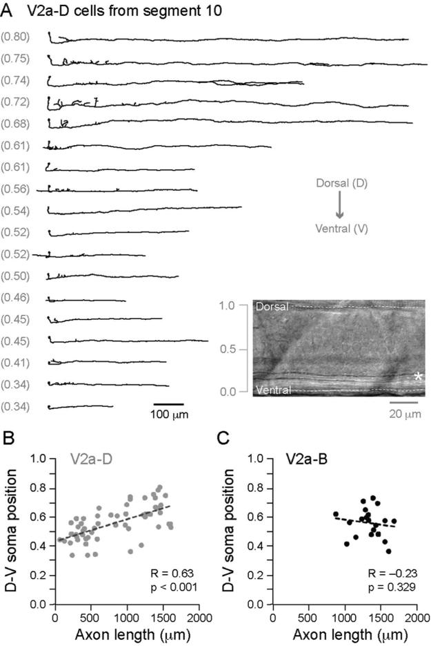
Figure 3. V2a-Ds have longer axons related to dorso-ventral soma position.
(A) Reconstructions of V2a-D cells arranged from top to bottom according to their dorso-ventral location (noted parenthetically to the left). All of the cells originate from body segment 10. Inset is a DIC image that illustrates how dorso-ventral location is normalized according to the top (1.0) and bottom (0.0) edges of spinal cord. Asterisk marks the large Mauthner axon.
(B) A comparison of dorso-ventral (D-V) soma position versus descending axon length reveals a significant relationship for V2a-Ds located between segments 5-15 (n = 57). Note, we limited our analysis to this region because V2a-Ds can extend up to 20 body segments (see Fig. 2D) and we did not want the end of the spinal cord (~segment 35) to act as a limiting factor.
(C) As in B, but no significant relationship is found for V2a-Bs (n = 20).

