
FIGURE 1.
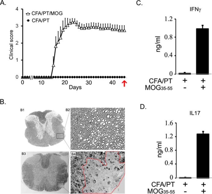
Characterization of clinical pathological state of EAE in chronic mouse model in B6. A, EAE was induced in C57B6 mice using MOG(35–55) peptide emulsified in CFA, and pertussis toxin was given on days 0 and 2 post-immunization. Clinical score was recorded daily (n = 10). The control group was given complete Freund's adjuvant/pertussis toxin without a peptide. On day 45, blood was drawn for analysis of isolated plasma. Red arrow indicates the time of sampling. B, at the end of the experiment, spinal cords were harvested from control (panels B1 and B2) and diseased mice (panels B3 and B4) to examine demyelination. Panels B1 and B3 show the whole mid-thoracic spinal cord section, and panels B2 and B4 show closer images from antero-lateral areas of the white matter. Completely normal and healthy axons can be appreciated on panel B2, and on panel B4 an area with clear demyelination is outlined in red. In addition, in areas that surround the demyelinating lesion, a patchy demyelination, dysmorphic and collapsed axons can be observed, too. C and D, recall response in cells isolated from lymph nodes, stimulated with 50 μg of MOG(35–55) for 72 h. Cell supernatant was used for measuring the levels of IFNγ and IL17 by ELISA (n = 4).