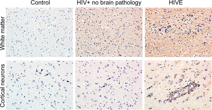
FIGURE 10.
DYRK1A expression in clinical samples. Representative images of DYRK1A immunolabeling in the white matter (original magnification ×20) and prefrontal cortex (original magnification ×40) of control brains, HIV+ without brain pathology, and HIVE (n = 4/group). A modest labeling of DYRK1A was observed in normal white matter and frontal cortex. Strikingly, intense labeling of axons and neuronal soma were observed in HIV+ without brain pathology, and this pattern was even stronger in HIVE cases. Arrows and arrowheads indicate neurons and glial cells, respectively.

