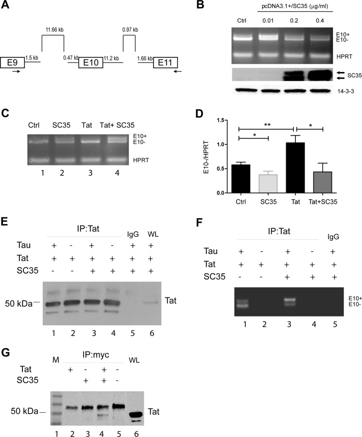
FIGURE 6.
Direct evidence that Tat promotes exon 10 exclusion. A, scheme of TAU minigene as reported previously (20, 52). B, titration of SC35 overexpression in 293T cells to determine the concentration of pSC35 at which E10+ and E10− TAU mRNA are equally produced (0.01 μg/ml) in the cells transfected for 36 h with the TAU minigene. HPRT was PCR-multiplexed and used as an internal control (Ctrl) and for normalization. Levels of SC35 upon its expression were detected by Western blots (middle panel). Anti-14-3-3 was used as a loading control. C, multiplex RT-PCR to detect E10+ and E10− TAU RNAs in controls, cells transfected with SC35, Tat, or both. HPRT was used for normalization. D, quantitatively, the TAU E10 exclusion is increased when Tat is present, and overexpression of SC35 prevents this up-regulation. One and two asterisks represent the significance between groups (p ≤ 0.05 and p ≤ 0.01, respectively) resulting from three independent experiments. E, Western blotting to detect Tat in cells transfected with pEYFP-Tat, Myc-SC35, and TAU minigene in the indicated combinations after immunoprecipitation (IP) with anti-Tat antibody or IgG control. WL stands for whole lysates and was used as a positive control for the expression of Tat. Note that EYFP-Tat is about 50 kDa. F, image of a representative agarose gel showing RT-PCR-amplified E10+ and E10− Tau RNAs in the indicated samples as in E. G, representative Western blots (n = 3) to detect Tat in 293T cells transfected with pEYFP-Tat101 (lane 2), Myc-SC35 (lane 3), co-transfected with Myc-SC35 and pEYFP-Tat101 (lane 4), and control mock-transfected (lane 5). M and WL stand for marker and whole lysate (lanes 1 and 6, respectively).

