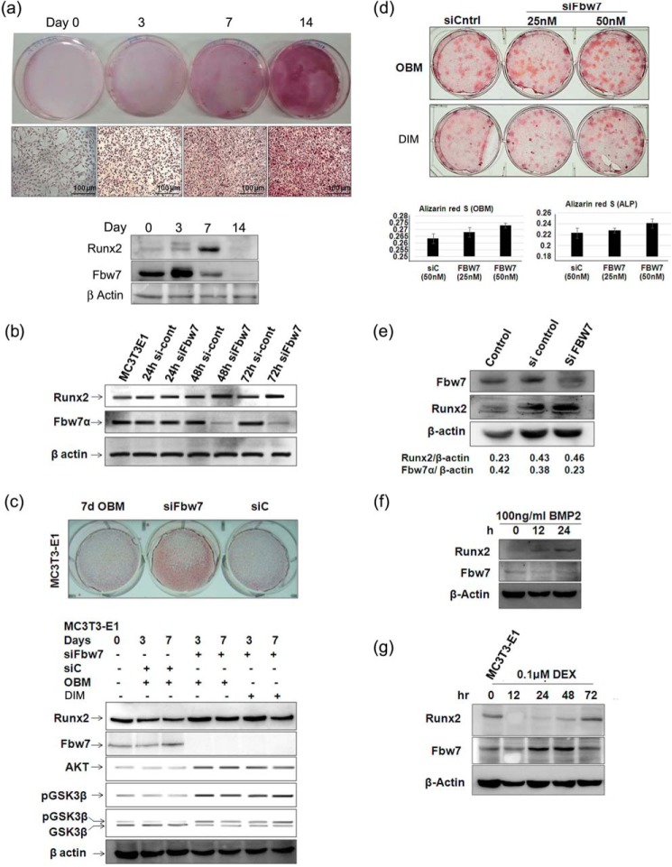
FIGURE 5.
Fbw7 knockdown restores Runx2 expression and enhances osteoblast differentiation. a, MC3T3-E1 cells were allowed to differentiate in DIM for 0, 3, 7, and 14 days. Following differentiation, Alizarin Red staining was performed after the indicated time points and visualized under a bright field microscope (20×; Leica) for analysis (upper panel). Whole cell lysates were prepared after 0, 3, 7, and 14 days followed by immunoblotting with anti-Runx2 and Fbw7α antibodies; β-actin was probed as a loading control (lower panel). b, lysates of MC3T3-E1 cells transfected with siControl (si-cont) or siFbw7α for 24, 48, and 72 h were immunoblotted with anti-Fbw7 and anti-Runx2 antibodies. β-Actin was probed as a loading control. c, MC3T3-E1 cells were transfected with either siControl (siC) or siFbw7α. 72 h after transfection, culture medium was changed every alternate day for 7 days. Cells were fixed and stained with Alizarin Red, and mineralized nodules were photographed (upper panel). MC3T3-E1 cells were transfected with siControl or siFbw7α. After 3 and 7 days, WCEs prepared were immunoblotted with anti-Fbw7 and anti-Runx2 antibodies. β-Actin was probed as a loading control (lower panel). OBM, osteoblast growth medium. d, primary osteoblast cells isolated from BALB/c mice calvaria were transfected with siControl or siFbw7α. 72 h after transfection, culture medium was changed alternatively for 14 days followed by Alizarin Red staining and imaging under a microscope. 1 ml of cetylpyridinium chloride was added to each well, readings were taken at 595 nm, and the graph was plotted. e, primary osteoblast cells isolated from BALB/c mice calvaria were transfected with siControl or siFbw7α. 72 h after transfection, and culture medium was changed every alternate days for 14 days followed by WCEs preparation and immunoblotting with anti-Fbw7 and anti-Runx2 antibodies. β-Actin was probed as a loading control. f, lysates of MC3T3-E1 cells treated with 100 ng/ml BMP2 and g, dexamethasone (DEX, 0.1 μm)-treated lysates were resolved on 10% SDS-PAGE and immunoblotted with anti-Runx2 and anti-Fbw7 antibodies. Data are representative of minimum three independent experiments.

