FIGURE 7.
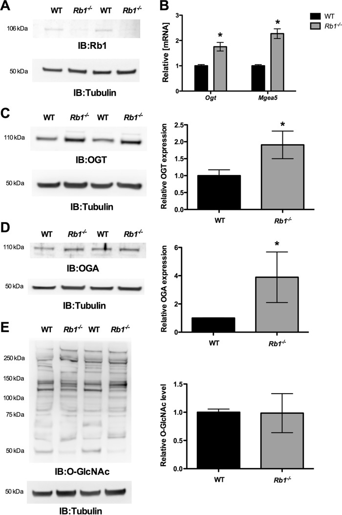
Loss of Rb1 increased Ogt and Mgea5 expression. MEFs that lack Rb1 were compared with the WT cells for the expression of Ogt and Mgea5. A, Western blot analysis (IB) showed significant loss of Rb1 in Rb1−/− cells compared with WT MEFs. B, significant increase in the Ogt (n = 4; p < 0.001) and Mgea5 (n = 4; p < 0.0001) mRNA level was observed in Rb1−/− MEFs. C and D, Western blot analysis showed a significant increase in the OGT (n = 3; p < 0.005) and OGA (n = 3; p < 0.05) protein level, whereas the O-GlcNAc level was not changed (E). Error bars, S.D. *, p < 0.05.

