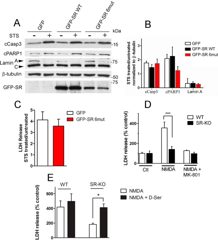
FIGURE 5.
Effect of SR translocation in cell death mechanisms. A, primary neuronal cultures obtained from SR-KO mice were infected at DIV5 with lentivirus harboring GFP, GFP-SR, or GFP-SR 6mut. On DIV10, neurons were treated with STS (1 μm) for 8 h in the presence of saturating levels of the NMDAR co-agonist glycine (50 μm). Total cell homogenates were probed for cleaved caspase 3, PARP-1, and lamin A cleavage by Western blot analysis. Lower panel depicts GFP-SR expression in total cell homogenate by anti-SR antibody. B, ratio of cleaved caspase3, PARP-1, and lamin A between STS-treated and -untreated cells normalized by β-tubulin levels. C, primary neuronal cultures obtained from SR-KO mice were infected at DIV5 with lentivirus harboring GFP or GFP-SR 6mut. On DIV10, neurons were treated with STS (1 μm) for 8 h in the presence of 30 μm MK-801 to evaluate cell death that was not mediated via NMDARs. The results are expressed as the ratio of LDH release by STS and untreated cultures. D, SR-KO cultures are less susceptible to NMDAR-mediated cell death. LDH release was monitored from cultures treated with no NMDA, 30 μm NMDA, or NMDA plus 20 μm MK-801. E, cell death was restored to WT levels when SR-KO primary neuronal cultures were supplemented with 10 μm d-serine. The results are mean ± S.E. of four (A–C and E) or nine (D) experiments with different culture preparations. *, ***, different from control at p < 0.05 and 0.001, respectively.

