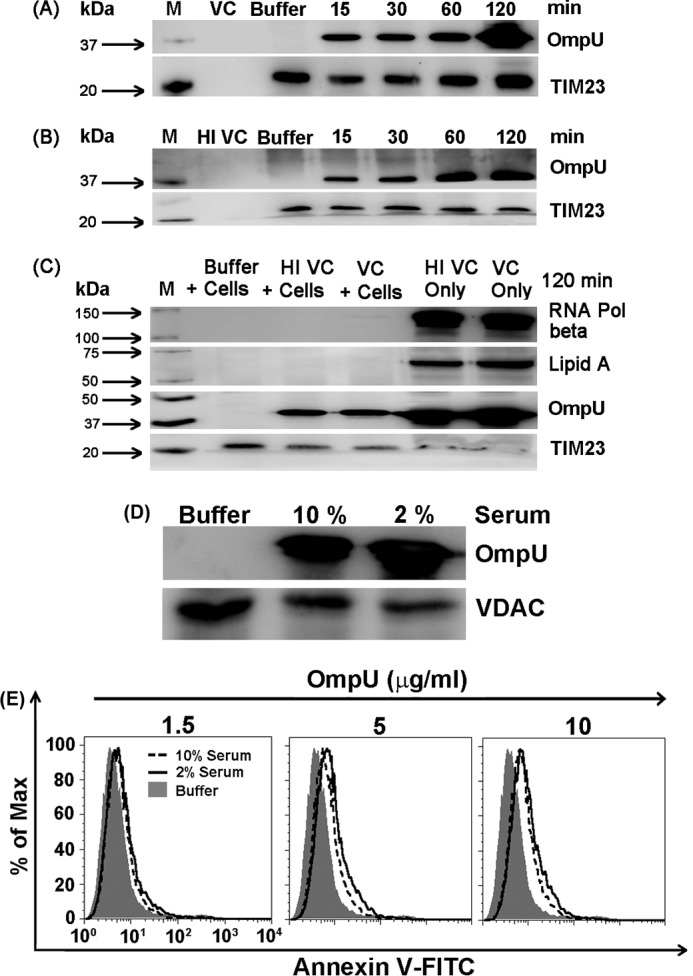
FIGURE 7.

OmpU is translocated to the mitochondria of target cells co-cultured with live and heat-inactivated V. cholerae. A, Western blots showing translocation of OmpU to the target cell mitochondria from live V. cholerae cells. THP-1 monocytes were infected with live V. cholerae (VC) at a multiplicity of infection of 10 and subjected to different incubation periods. PBS (Buffer)-treated cells and media containing only V. cholerae were used as controls. After respective incubations, mitochondrial fractions were isolated from all of the samples and analyzed for the presence of OmpU. The blots are representative of three or more independent experiments. B, Western blots showing translocation of OmpU to the host cell mitochondria from the heat-inactivated V. cholerae. THP-1 monocytes were infected with heat-inactivated V. cholerae (HI VC) at a multiplicity of infection of 10 for different incubation periods and analyzed for the presence of OmpU in a similar manner as mentioned above. The blots are representative of three independent experiments. C, control blot showing the absence of any bacterial component in purified mitochondria from co-culture of V. cholerae and mammalian cells. THP-1 monocytes were infected with live and heat-inactivated V. cholerae for 2 h. Mitochondrial lysates and bacterial cell lysates (live and heat-inactivated V. cholerae) were prepared and subjected to Western blot analysis for the detection of RNA polymerase beta (bacterial cytoplasmic marker) and lipid A, a component of LPS (bacterial outer membrane marker), along with OmpU and TIM23. D, an increase in serum concentration from 2 to 10% does not affect OmpU translocation to mitochondria. The Western blot shows mitochondrial translocation of OmpU to cells cultured in 2 and 10% serum-containing media. THP-1 monocytes were plated in 2 or 10% serum-containing medium, treated with 10 μg/ml OmpU, and incubated for 2 h. A mitochondrial fraction was isolated from all of the samples and analyzed for the presence of OmpU by Western blotting. Voltage-dependent anion channel (VDAC) was used as the mitochondrial marker and loading control. The blot is representative of two independent experiments. E, increase in serum concentration from 2 to 10% does not affect OmpU-induced PCD. A histogram overlay shows only the annexin V-positive population of OmpU-treated cells cultured in 2 and 10% serum-containing media. THP-1 monocytes cultured in 2 and 10% serum-containing media were treated with different doses of OmpU (1.5, 5, or 10 μg/ml) or buffer. Cells were harvested and stained with annexin V-FITC and PI. The histogram overlay represents a gated population comprising only annexin V-FITC single-positive cells. The plots are representative of three independent experiments.
