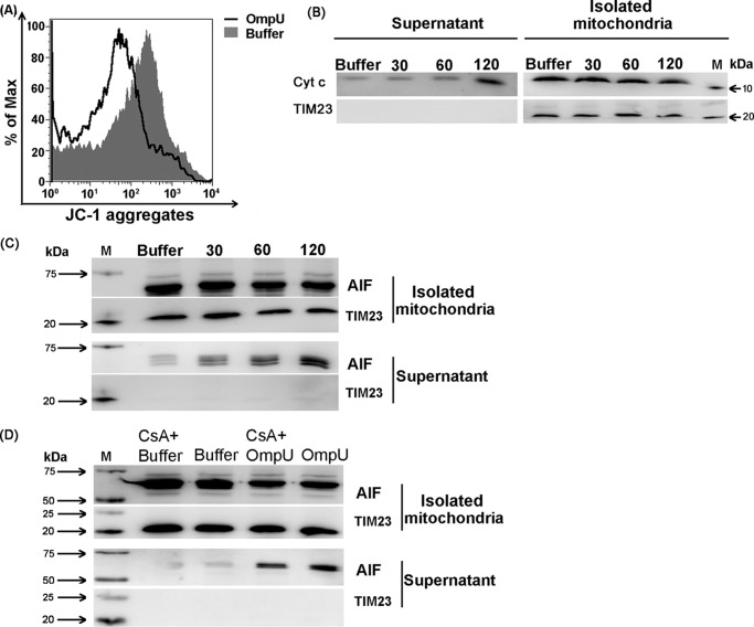
FIGURE 8.
OmpU directly induces loss of mitochondrial membrane potential and AIF release from freshly isolated mitochondria. A, histogram overlay represents the increase in loss of mitochondrial membrane potential in OmpU-treated mitochondria compared with buffer treatment. Mitochondria were isolated from 3 × 10⁷ THP-1 monocytes, and exactly equal aliquots of isolated mitochondria were incubated with 5 μg/ml OmpU or buffer for 30 min. Following incubation, mitochondria were stained with JC-1 dye. In the corresponding histogram overlay, the decrease in the red fluorescence of JC-1 aggregates gives a measure of decreased mitochondrial membrane potential in mitochondria. The plot is representative of three independent experiments. B and C, Western blots showing release of cytochrome c (B) and AIF (C) from the treated mitochondria and corresponding increase in the supernatant with an increase in incubation time after OmpU treatment. Equal aliquots of isolated mitochondria were treated with 5 μg/ml OmpU and incubated for different time periods or with buffer for 2 h. After the respective incubations, mitochondria were harvested, and supernatants were collected separately for each sample. Supernatants and mitochondria were analyzed for the presence of cytochrome c and AIF by Western blot analysis. The blots are representative of three or more independent experiments. D, Western blots showing inhibition of AIF release from isolated mitochondria by CsA. Freshly isolated mitochondria were pretreated with 1 μm CsA for 30 min, followed by treatment with 5 μg/ml OmpU for 90 min. Following incubation, samples were subjected to Western blotting as described above. Blots are representative of three independent experiments.

