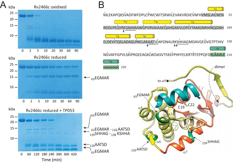
FIGURE 3.
Limited proteolysis experiments show significant conformational differences between the redox forms of Rv2466c. A, SDS-PAGE showing the trypsin cleavage profile for Rv2466cOX, Rv2466cRED, and Rv2466cRED in the presence of TP053. B, the peptide bonds that are cleaved specifically by trypsin are shown with arrows. The N-terminal sequences of selected proteolytic fragments are underlined in the amino acid sequence and labeled in dark green in the schematic representation of Rv2466c.

