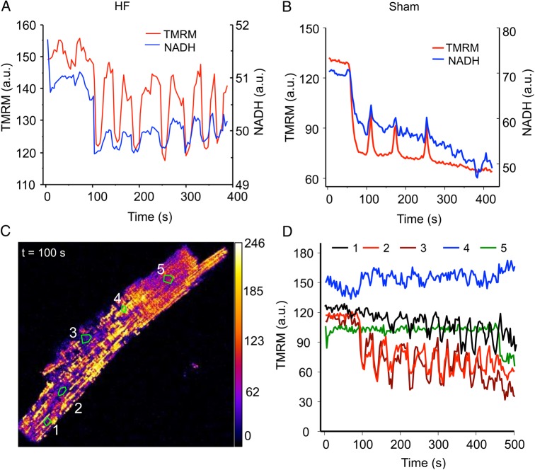
Figure 3.
Uncoupled laser flash-induced mitochondrial network oscillations in HF cardiomyocytes. (A) Oscillations of an HF cardiomyocyte involving mitochondrial membrane potential (TMRM) and NADH; (B) oscillations of a sham cell; (C) the snapshot of an oscillating HF cardiomyocyte showing heterogeneous mitochondrial energetic state; and (D) plots of time profile of membrane potential (measured by TMRM) of various mitochondrial clusters (curves 1–5 represent zones 1–5 marked in C). A total of 30 cells from 5 sham hearts and 40 cells from 7 HF hearts were examined.

