Figure 3.
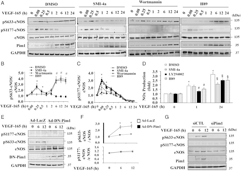
VEGF induces Pim1 expression and sustained phosphorylation of eNOS at Ser-633 in HUVECs. (A) HUVECs were pre-treated with either DMSO or Pim1 inhibitor SMI-4a (10 µmol/L), PI3K inhibitor wortmannin (100 nmol/L), and PKA inhibitor H89 (10 µmol/L) for 2 h and then stimulated with 50 ng/mL VEGF-165 for different time points as indicated. Phosphorylation of eNOS and expression of eNOS and Pim1 were then analysed by western blot. (B) Quantitative analysis phosphorylation of Ser-633-eNOS (n = 4). *P < 0.05 compared with H89 at indicated time points; #P < 0.05 compared with SMI-4a at indicated time points by two-way ANOVA. (C) Quantitative analysis phosphorylation of Ser-1177-eNOS (n = 4). *P < 0.05 compared with LY294002 at indicated time points by two-way ANOVA. (D) NO in cell culture medium at 1 and 24 h after addition of VEGF was analysed and normalized to VEGF 0 h/DMSO group (n = 4). *P < 0.05 compared with DMSO at 0 h; #P < 0.05 vs. DMSO at 1 h after VEGF stimulation; §P < 0.05 vs. DMSO at 24 h after VEGF stimulation (two-way ANOVA). (E) DN-Pim1 attenuates VEGF-induced eNOS phosphorylation at Ser-633. HUVECs were transduced with either Ad-Lacz or Ad-DN-Pim1 virus (50 MOI). 48 h after transduction, cells were then stimulated with 50 ng/mL VEGF-165 for 6 and 12 h. The phosphorylation and expression of eNOS were determined by western blot. (F) Quantitative analysis of p-eNOS/total eNOS ratio (n = 4). *P < 0.05 compared with Ad-DN-Pim1 by two-way ANOVA. (G) 72 h after transfection of either Pim1 siRNA (siPim1) or control siRNA (siCTL), cells were then stimulated with 50 ng/mL VEGF-165 for 6 and 12 h. The eNOS phosphorylation was determined by western blot (n = 4).

