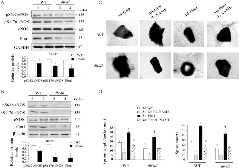
Figure 6.
Overexpression of Pim1 ameliorates impairment of angiogenesis in ex vivo. (A) and (B) Hearts and aortas were harvested from control C57BLKS/J mice (WT) and db/db mice. eNOS phosphorylation and expression of eNOS and Pim1 were analysed by western blot. *P < 0.01 compared with WT mice (Student's t-test), n = 6. (C) Aortic rings were incubated with Ad-Pim1 and Ad-GFP in opti-MEM medium for 24 h and then embedded in Matrigel. As microvessels began to branch and develop, some wells were pre-treated with 0.5 mmol/L l-NAME (n = 4 per group). Phase-contrast photos of individual explants were taken using EVOS® FL Cell Imaging System. (D) Analysis of the microvessel growth (sprout length and sprout number) was performed by image J program. *P < 0.05 compared with WT/Ad-GFP group; #P < 0.05 compared with WT/Ad-Pim1 group; §P < 0.01 compared with either db/db/Ad-GFP group or db/db/Ad-Pim1/L-NAME group (two-way ANOVA). n = 4. Scale bars = 2000 μm.

