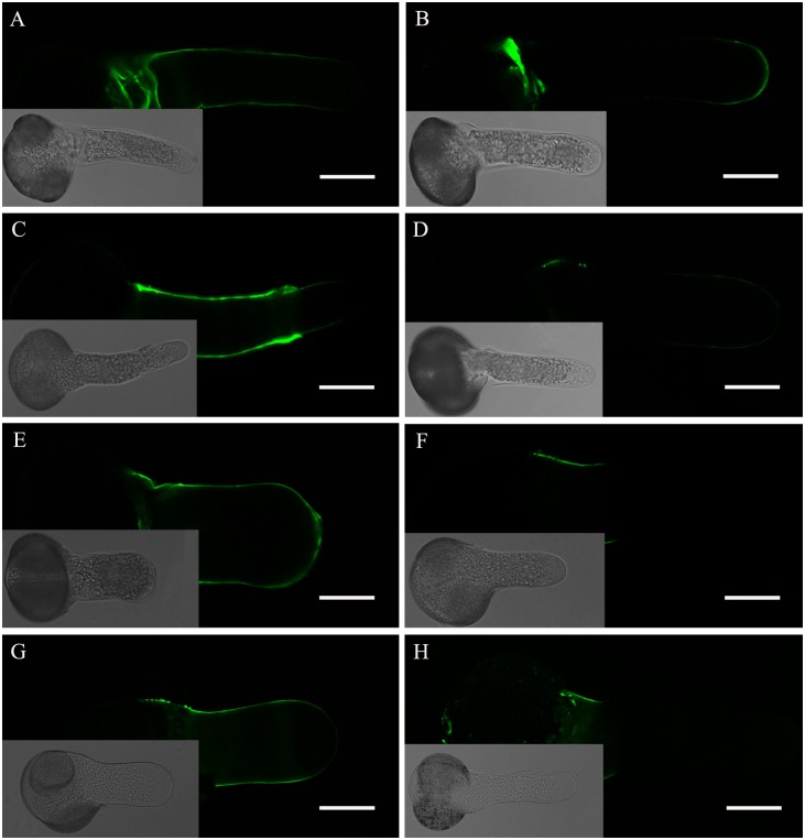
Fig 6. Fluorescent immunolabeling of pectins in a Picea wilsonii pollen tube.
(A) JIM5 labeling of pollen tubes cultured in standard medium. Strong fluorescence was detected along the entire tube shank wall, excluding the tip. (B) JIM7 labeling of pollen tubes cultured in standard medium. Esterified pectins were found at the tip of the pollen tube. (C) JIM5 labeling of pollen tubes treated with medium containing 0.2% DMSO. The result is consistent with that obtained for the control (A). (D) JIM7 labeling of pollen tubes treated with medium containing 0.2% DMSO. The distribution of esterified pectins is consistent with that seen in the control (B). (E) JIM5 labeling of pollen tubes cultured in the presence of 0.5 μM TSA. The fluorescence signal was distributed along the pollen tube wall. (F) JIM7 labeling of pollen tubes incubated in medium containing 0.5 μM TSA. Esterified pectins were found only in the basal part of the pollen tube wall. (G) JIM5 labeling of pollen tubes cultured in the presence of 0.5 mM NaB. (H) JIM7 labeling of pollen tubes incubated in medium containing 0.5 mM NaB. Bar = 50 μm.

