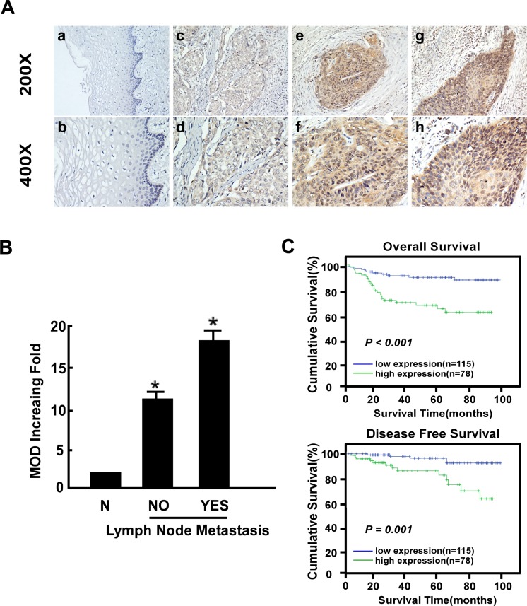
Fig 3. Immunohistochemical detection of B3GNT3 protein expression in paraffin-embedded tissues.
Positive B3GNT3 staining was observed mainly in the cytoplasm of cervical cancer cells. (A) a and b, B3GNT3 expression was not detected in normal cervical tissues; c and d, representative images of weak B3GNT3 staining in cervical cancer tissues; e and f, representative images of moderate B3GNT3 staining in cervical cancer tissues; g and h, representative images of strong B3GNT3 staining in cervical cancer tissues. (B) The statistical analyses of the average mean optical density (MOD) of B3GNT3 staining in the lymph node metastasis group and the lymph node metastasis-free group. *P < 0.05. (C) Kaplan-Meier curves of univariate analysis data (log-rank test).The overall survival (OS) and disease-free survival (DFS) for the patients with high versus low B3GNT3 expression.

